Paging Dr. Offit: Robert F. Kennedy is NOT Lying: Everyone Knows that Thimerosal Causes Neuroinflammation. Score: Kennedy +3, Offit, -3. Did Offit Even Read Burbacher?
It's utterly frightening that someone in Offit's position is so unfamiliar with the science that shows that thimerosal causes neuroinflammation. Here's another reading list for Offit.
In his Substack, Vaccine Activist Paul Offit claimed Mr. Kennedy lied about neuroinflammation caused by thimerosal.
He also claimed that mercury from thimerosal does not cross the blood/brain barrier while citing a study (Burbacher et al.) that shows not only does mercury from thimerosal cross the blood/brain barrier - it lasts so long in the brain with such low clearance that, unlike mercury from ethylmercury, it was not possible to calculate a clearance rate.
Offit refuses to read the scientific literature that the rest of us have read and are quite familiar with.
So I provide Dr. Offit with yet another reading list.
Offit: “Dr. Paul Offit Issues a Rebuttal to Robert F. Kennedy's Joe Rogan Podcast Saying Thimerosal Can Cross the Blood Brain Barrier, But It Is Not Harmful
"Unlike inorganic mercury, methylmercury can cross cell membranes and do harm...Ethylmercury (thimerosal), however, is not methylmercury. While methylmercury has a half-life in the bloodstream of about 70 days, ethylmercury has a half-life of seven days. So, it’s much less likely to accumulate and do harm...It was not surprising that Thomas Burbacher found trace quantities of ethylmercury in the brain of infant monkeys injected with thimerosal, which can cross cell membranes. But RFK Jr. lied when he said that the mercury had caused 'severe inflammation.'"
From the Burbacher study, which Offit claims to have read and understood:
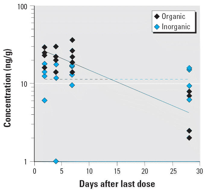
“A plot of the organic and inorganic Hg concentrations in the brain of thimerosal-exposed infant monkeys sacrificed at various times during the washout period is shown in Figure 7. There was a significant decrease in organic Hg in the brain over the washout period (p < 0.01). The apparent T1/2 for the washout of organic Hg from the brain was 14.2 ± 5.2 days, which is significantly shorter than the T1/2 for total Hg in brain (p < 0.01). The inorganic form of Hg was readily measurable in the brain of the thimerosal-exposed monkeys. The average concentration of inorganic Hg did not change across the 28 days of washout and was approximately 16 ng/mL (Figure 7). This level of inorganic Hg represented 21–86% of the total Hg in the brain (mean ± SE, 70 ± 4%), depending on the sacrifice time. These values are considerably higher than the inorganic fraction observed in the brain of MeHg monkeys (6–10%).” (emphasis added for Dr. Offit’s benefit).
Here is Figure 7. First, if Offit was right about mercury from thimerosal not being able to cross the blood-brain barrier, this figure would not exist. The value on the y-axis, brain tissue concentration (ng/g) would be zero from Day 1.
Worse, people with autism have altered blood/brain barriers. Maybe read something somtime, Paul? Especially when infant brains are at stake? (Here’s a study LINK)
Score: Kennedy 1, Offit, -1
The blue diamonds represent the inorganic form of mercury from thimerosal. See how flat the line is? No clearance. That’s far worse than methylmercury, which is shown in another Figure in the Burbacher et al. paper.
Score: Kennedy 2, Offit, -2.
Now, remember the claim that Kennedy lied about neuroinflammation caused by thimerosal?
Burchacher cited Vargas et al, pointing to the fact of chronic neuroinflammation in autism. Rather than connect the dots, or even show ANY concern over neuroinflammation in the brains of kids with autism, Offit wants to claim Kennedy lied. Guess what? Bobby has read just about everything on Thimerosal and knows it causes neuroinflammation. I even included a link to the Burbacher study, which Offit clearly has not read.
See the short reading list below. Guess what?
Score: Kennedy +3, Offit, -3
Dr. Offit’s Reading List:
Abstract. Evidence suggests that the effect of heavy metals on neuroimmune cells lead to neurogenic inflammatory responses. In this study, immune cells [mast cells (MCs) and microglia] and pro-neuroinflammation cytokines (interleukin-1b and tumor necrosis factor-α) were assessed in the prefrontal lobe of rat brains exposed to thimerosal in different timeframes. A total of 108 neonatal Wistar rats were divided into three groups having three subgroups. The experimental groups received a single dose of thimerosal (300 μg/kg) postnatally at 7, 9, 11, and 15 days. The vehicle groups received similar injections of phosphate-buffered saline in a similar manner. The control groups received nothing. Samples of the prefrontal cortex were collected and prepared for stereological, immunohistochemical, and molecular studies at timeframes of 12 or 48 h (acute phase) and 8 days (subchronic phase) after the last injection. The average density of the microglia and MCs increased significantly in the experimental groups. This increase was more evident in the 48 h group. At 8 days after the last injection, there was a significant decrease in the density of the MCs compared to the 12 and 48 h groups. Alterations in pro-inflammatory cytokines were significant for all timeframes. This increase was more evident in the 48 h group after the last injection. There was a significant decrease in both neuroinflammatory cytokines at 8 days after the last injection. It was found that ethylmercury caused abnormal neurogenic inflammatory reactions and alterations in the neuroimmune cells that remained for a longer period in the brain than in the blood.
Dórea JG. Integrating experimental (in vitro and in vivo) neurotoxicity studies of low-dose thimerosal relevant to vaccines. Neurochem Res. 2011 Jun;36(6):927-38. doi: 10.1007/s11064-011-0427-0. Epub 2011 Feb 25. PMID: 21350943.
To the point of include Ijaz, in case you cannot connect dots: Human tissues have a regular pattern of expressed proteins involved in inflammation. Here is the signature in kidney damage.
Ijaz MU, Majeed SA, Asharaf A, Ali T, Al-Ghanim KA, Asad F, Zafar S, Ismail M, Samad A, Ahmed Z, Al-Misned F, Riaz MN, Mahboob S. Toxicological effects of thimerosal on rat kidney: a histological and biochemical study. Braz J Biol. 2021 Aug 27;83:e242942. doi: 10.1590/1519-6984.242942. PMID: 34468508.
Thimerosal is an organomercurial compound, which is used in the preparation of intramuscular immunoglobulin, antivenoms, tattoo inks, skin test antigens, nasal products, ophthalmic drops, and vaccines as a preservative. In most of animal species and humans, the kidney is one of the main sites for mercurial compounds deposition and target organs for toxicity. So, the current research was intended to assess the thimerosal induced nephrotoxicity in male rats. Twenty-four adult male albino rats were categorized into four groups. The first group was a control group. Rats of Group-II, Group-III, and Group-IV were administered with 0.5µg/kg, 10µg/kg, and 50µg/kg of thimerosal once a day, respectively. Thimerosal administration significantly decreased the activities of catalase (CAT), superoxide dismutase (SOD), peroxidase (POD), glutathione reductase (GR), glutathione (GSH), and protein content while increased the thiobarbituric acid reactive substances (TBARS) and hydrogen peroxide (H2O2) levels dose-dependently. Blood urea nitrogen (BUN), creatinine, urobilinogen, urinary proteins, kidney injury molecule-1 (KIM-1), and neutrophil gelatinase-associated lipocalin (NGAL) levels were substantially increased. In contrast, urinary albumin and creatinine clearance was reduced dose-dependently in thimerosal treated groups. The results demonstrated that thimerosal significantly increased the inflammation indicators including nuclear factor kappaB (NF-κB), tumor necrosis factor-α (TNF-α), Interleukin-1β (IL-1β), Interleukin-6 (IL-6) levels and cyclooxygenase-2 (COX-2) activities, DNA and histopathological damages dose-dependently. So, the present findings ascertained that thimerosal exerted nephrotoxicity in male albino rats.
So, are the hallmarks of thimerosal-induced inflammation - nuclear factor kappaB (NF-κB), tumor necrosis factor-α (TNF-α), Interleukin-1β (IL-1β), Interleukin-6 (IL-6) levels and cyclooxygenase-2 (COX-2) over-expressed in the brains of people with autism?
Let’s find out. (These are just a subsample of the ample evidence available via Pubmed):
NF-κB: YES
Tumor necrosis factor-α (TNF-α
IL-6: YES
Cox2: YES


Despite some hiccups, I'm very grateful this subject is now being openly discussed. It feels like progress to me :)
Dr. Profit already had this discussion years ago with Mr. Kennedy and had to admit he was wrong. But that didn’t stop Dr. Profit from continuing to lie about it. Remember that initially he was afraid of the COVID shots because, as he said, mRNA tech never worked before and might make people look at the entire vax schedule. Glad he’s getting what he feared. Most of our childrens problems and many of the adult chronic degenerative diseases are from those vaxs. Read the inserts they never provide you. There is no real informed consent.